Microscope Magnification: A Simple Calculation

The concept of microscope magnification is fundamental to the field of microscopy, playing a pivotal role in scientific research, medical diagnostics, and various industrial applications. Understanding how to calculate microscope magnification is not only essential for those using microscopes but also for anyone interested in the intricacies of the microscopic world.
Understanding Microscope Magnification

Magnification, at its core, is the process of enlarging an object to make its details more visible. In the context of a microscope, it refers to the ability to enlarge the image of a specimen so that its minute structures can be observed. This process is achieved through a combination of optical lenses and precise adjustments.
The significance of microscope magnification lies in its ability to reveal the unseen. It allows scientists and researchers to explore the microcosm, studying cells, tissues, and organisms in unprecedented detail. From identifying pathogens in medical samples to analyzing the intricate patterns of minerals, microscope magnification is a powerful tool for knowledge acquisition and problem-solving.
The Fundamentals of Microscope Magnification

Magnification in microscopy is a product of two primary components: the objective lens and the eyepiece. The objective lens, positioned closest to the specimen, is responsible for the initial magnification. It collects light from the specimen and forms a real image, which is then further magnified by the eyepiece.
The objective lenses come in various magnifications, typically ranging from 4x to 100x. A 4x objective, for instance, will enlarge the specimen by four times its original size, making it four times larger and easier to observe. The eyepiece, on the other hand, often has a fixed magnification of 10x. This means that it will further enlarge the image produced by the objective lens, resulting in a final magnification that is the product of the objective and eyepiece magnifications.
For example, if you use a 40x objective lens (4x magnification) and a 10x eyepiece, the final magnification would be 400x (40x x 10x). This means the specimen would appear 400 times larger than its actual size, revealing fine details that are otherwise invisible to the naked eye.
Practical Considerations for Microscope Magnification
When working with microscopes, it’s essential to consider the practical implications of magnification. While higher magnifications offer greater detail, they also come with certain limitations. One key consideration is the field of view, which decreases as magnification increases. This means that at higher magnifications, you will see a smaller portion of the specimen.
Additionally, the resolution of the microscope becomes a critical factor. Resolution refers to the microscope's ability to distinguish between two closely spaced objects. As magnification increases, the resolution must also increase to maintain clear and sharp images. This is where the quality of the microscope's lenses and optical system comes into play.
Another practical aspect is the depth of field. Higher magnifications often result in a shallower depth of field, which means that only a thin slice of the specimen will be in focus at any given time. This can make focusing and observing three-dimensional specimens more challenging.
Calculating Microscope Magnification
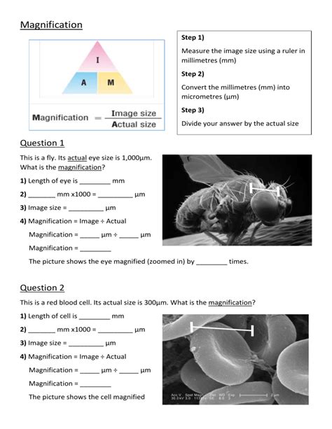
Calculating microscope magnification is straightforward once you understand the basic principles. As mentioned earlier, the final magnification is simply the product of the objective lens magnification and the eyepiece magnification. Mathematically, it can be represented as:
Final Magnification = Objective Lens Magnification x Eyepiece Magnification
Let's illustrate this with a few examples. Suppose you have a microscope with a 4x objective lens and a 10x eyepiece. The final magnification would be:
Final Magnification = 4x x 10x = 40x
Now, consider a microscope with a 100x objective lens and the same 10x eyepiece. The final magnification in this case would be:
Final Magnification = 100x x 10x = 1000x
It's important to note that while higher magnifications offer more detail, they also require more precise adjustments and may introduce challenges in terms of resolution and depth of field.
Real-World Applications and Implications

The practical applications of microscope magnification are vast and impactful. In medical laboratories, for instance, high-magnification microscopes are used to diagnose diseases by examining blood samples, tissue cultures, and even individual cells. This level of detail is crucial for identifying pathogens, detecting abnormalities, and determining the most effective treatments.
In materials science and engineering, microscopes are employed to analyze the microstructure of materials. This includes examining the grain structure of metals, identifying defects in semiconductor devices, and studying the morphology of polymers. The ability to magnify these materials provides insights into their mechanical properties, electrical behavior, and overall performance.
Moreover, in fields like environmental science and biology, microscopes are indispensable for studying microorganisms, aquatic organisms, and plant tissues. The magnification capabilities of these instruments enable researchers to study the behavior, morphology, and interactions of these organisms, contributing to our understanding of ecosystems and biological processes.
| Objective Lens Magnification | Eyepiece Magnification | Final Magnification |
|---|---|---|
| 4x | 10x | 40x |
| 10x | 10x | 100x |
| 40x | 10x | 400x |
| 100x | 10x | 1000x |

Frequently Asked Questions
How does microscope magnification impact image quality?
+
Higher magnifications can reveal more detail, but they also demand higher resolution from the microscope’s optical system. If the resolution is insufficient, images may appear blurry or distorted, despite the increased magnification.
Can I use different combinations of objective lenses and eyepieces to achieve specific magnifications?
+
Absolutely! Microscopes often come with a range of objective lenses and eyepieces, allowing users to customize the magnification to suit their specific needs. For instance, you could use a 20x objective lens with a 5x eyepiece to achieve a final magnification of 100x.
What are some common challenges associated with high magnification microscopy?
+
High magnifications often result in a shallower depth of field, making it challenging to keep the entire specimen in focus. Additionally, higher magnifications may require more precise adjustments and can introduce challenges related to lighting and contrast.
How can I choose the right magnification for my microscope observations?
+
The choice of magnification depends on the specific requirements of your observation. Lower magnifications provide a broader field of view and are suitable for initial specimen identification. Higher magnifications are ideal for detailed analysis and examination of fine structures. It’s often beneficial to start with lower magnifications and gradually increase as needed.
Are there any safety considerations when working with high-magnification microscopes?
+
Yes, safety is crucial when working with microscopes, especially at high magnifications. Always ensure proper handling of the microscope to avoid accidents. Additionally, be cautious when dealing with potentially hazardous samples, and follow laboratory safety guidelines.


